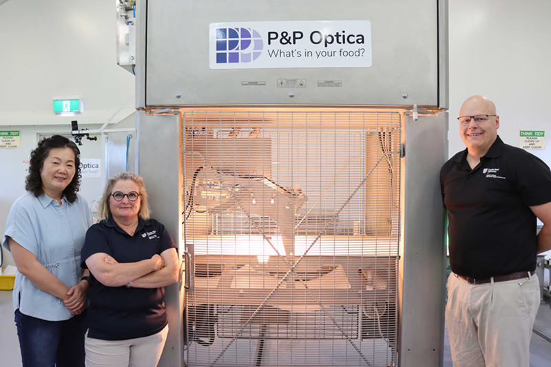
 Lihong Zheng, Jane Quinn and Stephen Cork in front of P&P Optica’s offal disease detection device at CSU.
The animal health clues hidden inside the carcase
For decades, disease detection in offal has been dependent on visual assessments by meat inspectors – a labour-intensive process that can vary between people and processing sites – but that's set to change with new technology.
To bring greater consistency and accuracy to the task, the new technology has been developed by Charles Sturt University (CSU) and P&P Optica with support from MLA and Tey’s Australia.
It works alongside meat inspectors to provide an objective and repeatable method of diagnosing disease and defects in offal.
The MLA Donor Company-funded project is investigating how hyperspectral imaging and machine learning can support more consistent disease assessment in lungs, livers and kidneys, while generating detailed health data that can be fed back to producers.
The goal of this initiative is to improve consistency, accuracy and decision making across the supply chain.
The brains behind the initiative
Leading the project is Professor Jane Quinn from CSU’s School of Agricultural, Environmental and Veterinary Sciences, who developed the original concept after identifying a need for more objective disease assessment at slaughter.
“The project itself builds on earlier proof-of-concept work we completed, showing machine learning could successfully differentiate between disease states in beef lungs,” she said.
“The idea first emerged almost a decade ago when we began exploring how technology could help overcome some of the limitations associated with traditional visual inspection and consistency of data capture.”
According to Jane, while meat inspectors play a critical role in protecting food safety and identifying the presence of disease in tissues destined for human or animal consumption, there can be limitations in the amount of information captured and reported.
“The role of the meat inspector is to identify whether the presence of disease that impacts food safety exists, not to necessarily diagnose the disease or defect and communicate it back to producers,” she said.
“You can look at a lot of animals from the outside and they all look the same, but the inside will tell a different story.
“This leaves a gap in the flow of information through the supply chain, which presented to us as an opportunity to develop a tool that could operate alongside existing inspection systems, and provide producers with consistent feedback to support better on-farm decision making.”
Machine learning
While Jane identified the industry challenge, turning veterinary knowledge into something a machine could understand was undertaken by Professor Lihong Zheng from CSU’s School of Computing, Mathematics and Engineering.
She worked in conjunction with P&P Optica’s model development team.
As a specialist in image processing and machine learning, Lihong focused on developing the algorithms capable of detecting and accurately diagnosing disease and defects in offal via camera.
At the same time, P&P Optica developed similar algorithms for hyperspectral image capture.
“My role in this project was to interpret Jane’s expert knowledge into machine-actionable information,” Lihong said.
“In simple terms, that meant teaching a computer to recognise the same disease indicators that an experienced meat inspector or veterinarian would identify during an assessment.”
Drawing on thousands of images collected throughout the project, P&P Optica trained machine learning models to distinguish between healthy tissue and six different disease conditions affecting lungs, livers and kidneys.
This included:
- liver abscess
- liver fluke
- kidney cysts
- pneumonia
- pleurisy
- nephritis.
The addition of hyperspectral imaging added another layer of information, allowing the system to analyse characteristics beyond those visible to the human eye.
“P&P Optica was then able to integrate the established machine learning model into a commercial product that was installed at CSU’s Food Pilot Plant for pre-commercial testing,” Lihong said.
“For the first time, we were able to see the technology operating under realistic processing conditions, assessing offal in real time and generating objective defect identification.
"In simple terms, that meant teaching a computer to recognise the same disease indicators that an experienced meat inspector or veterinarian would identify during an assessment.
“From there, the focus shifted to refining the system and validating its accuracy under commercial conditions.”
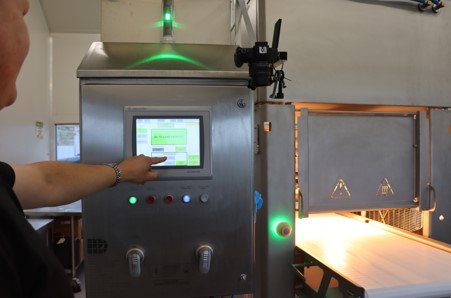
Dr Stephen Cork demonstrates how the offal disease detection device is operated. Image supplied.
Closing the feedback loop
On the processing floor, Dr Stephen Cork – a research fellow at CSU with extensive experience in meat processing and quality assurance – took the lead in ensuring the technology could perform reliably in a commercial environment.
“We were able to achieve an accuracy rating of 88% for our six identifiable diseases across the three offals – that will gradually improve as we gain access to more samples for scanning,” he said.
For Stephen, the significance of those results lies in the quality of information that can be returned to producers.
“You can look at a lot of animals from the outside and they all look the same, but the inside will tell a different story,” he said.
“By providing objective and consistent health data, the technology has the potential to give producers greater visibility of the health challenges affecting their livestock.
"This will help them identify trends, assess the effectiveness of management practices, and make more informed decisions to improve productivity and carcase quality over time.”
The next phase of the project will focus on refining the design of the scanning system by developing a more modular structure that can be seamlessly integrated into existing processing plant infrastructure and adopted more broadly across the red meat industry.